Hematologic tests are common and indispensable for clinical diagnosis. Among them, routine white blood cell (WBC) differential counts provide fundamental results for patients with infection, inflammation or leukocyte-related diseases. In modern hematology laboratory, the blood samples were initially examined with automated hematology analyzer by which the questionable results or unexpected flags are alarmed to the medical technologists who can recheck the blood smear manually. Traditionally, the accuracy and reliability of WBC differential count rely on experienced medical technologists under microscopic observation, therefore, the well-training, visual acuity, and adequate experience of medical technologists are very important.
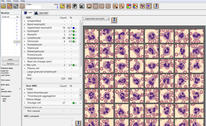
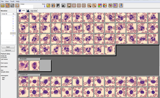
Since December 2018, the NCKUH Hematology laboratory adopted the digital microscopy system (CellaVision DM9600) which represents the most popular computer-aided instrument assisting WBC classification in the clinical hematology laboratory, including ID identification of blood smear by slide barcode, location of WBC area under low power lens, capture of WBC morphology with camera system under high power lens, and finally differentiation and counting of WBC according to the cell size, granules and nucleus etc.
This image system is differs from those of the past when only blood smears could be retained for cell morphology databank. More than 1000 blood smears are usually generated in the hematology laboratory each day and a lot of time is spent in looking for slides for pathological interpretation, case discussion, and clinical studies. In addition, blood smear slides can not only be stored for an unlimited time. The use of digital images can reduce eyestrain from prolonged periods of microscope viewing and improve test report efficacy and quality and expedite the integrity of data storage and retrieval Overall, the interpretation and retention of digital WBC images has largely increased the convenience of smear review and clinical communication.

