Integrating AI-Assisted Technologies into Prostate Cancer Diagnosis and Biopsy Workflows: From Physician Training to Clinical Decision-Making
KUN-CHE LIN, Attending Physician, Department of Urology
Prostate cancer has become the third most common cancer among men in Taiwan. Clinical diagnosis relies heavily on multi-parametric MRI (multiparametric magnetic resonance imaging; mpMRI) and the PI-RADS scoring system. However, image interpretation is often subjective, and the complexity of MRI-ultrasound fusion biopsy leads to a steep learning curve for junior physicians, hindering diagnostic efficiency and optimal resource allocation. The team developed an AI-powered Prostate Image Analysis System based on the U-Net3+ deep learning architecture. This system automates prostate segmentation, lesion detection, and PI-RADS scoring (average processing time: 52 seconds). Furthermore, we integrated the SECI knowledge creation model with Augmented Reality (AR) technology to project AI-generated 3D lesion models onto physical training phantoms, establishing a standardized curriculum from image interpretation to surgical simulation.
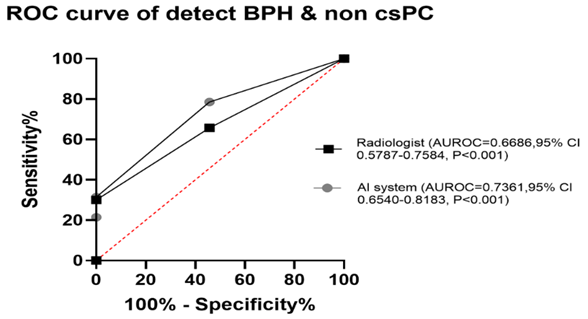
Enhanced Training Quality: Trainees demonstrated significantly improved accuracy in PI-RADS scoring and higher surgical confidence post-training (p-value < 0.05). Average operative time decreased annually; senior residents successfully reduced surgical duration to under 100 minutes (p-value < 0.0001). Through strategic alliances and physician-led lesion contouring, waiting times for mpMRI scans and lesion labeling were markedly reduced. The AI system achieved an AUC of 0.7361 in differentiating benign hyperplasia from low-grade cancer, outperforming radiologists’ area under the curve (AUC: 0.6686), thereby preventing unnecessary invasive biopsies. This project successfully integrates AI diagnostics with AR-assisted simulation. It not only enhances clinical quality and resource efficiency but also advances the implementation of precision medicine, serving as a model for digital transformation in prostate cancer diagnostic workflows.
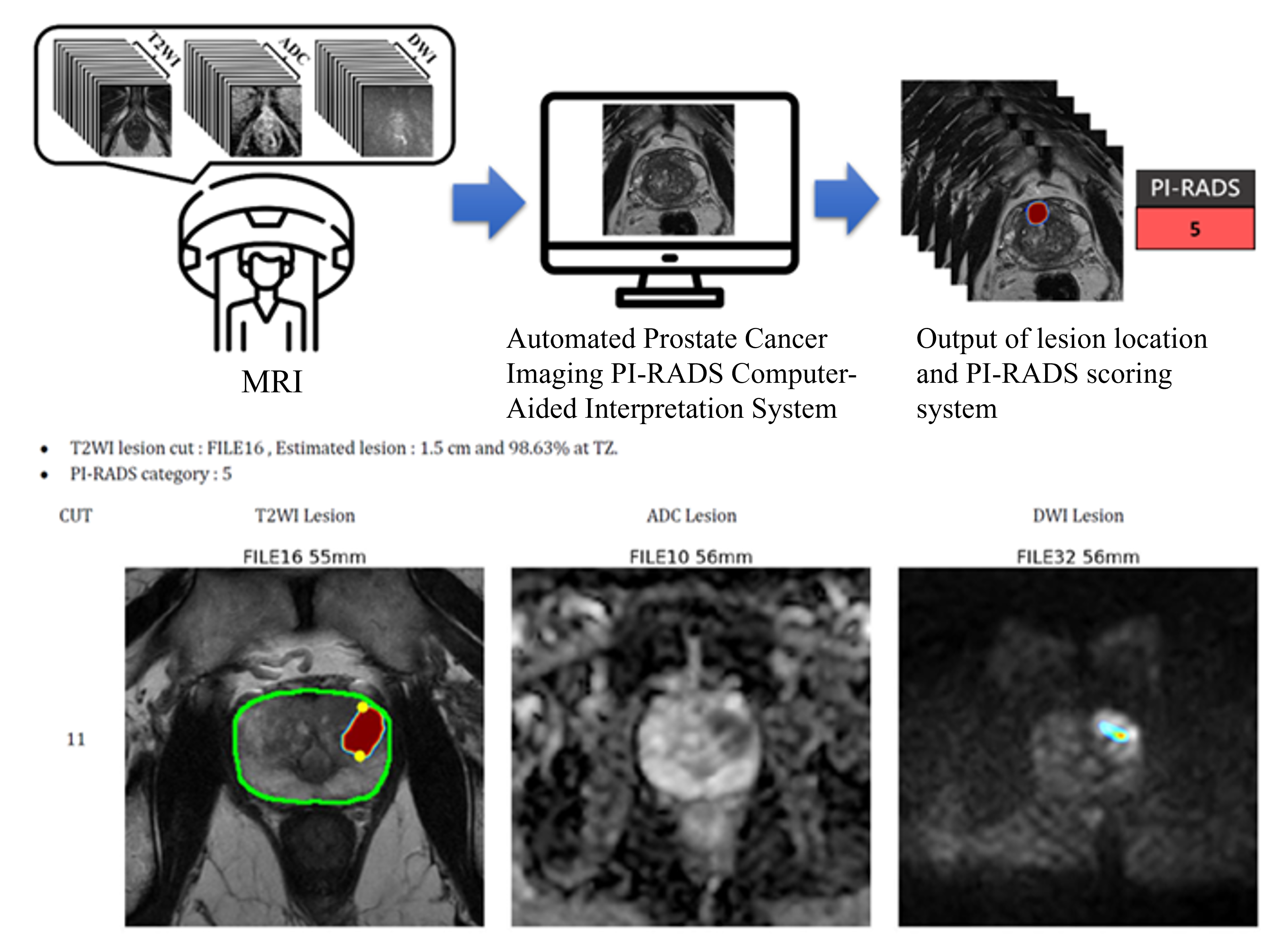
Figure 1. The artificial intelligence system analyzes the prostate mpMRI systemic flow chart and case demonstration.