
Automated Interpretation and Counting System for Cells in Bone Marrow Smears: A Wonderful Assistant for Hematologists
Ya-Ting Hsu, MD, Division of Hematology, Dept. of Internal Medicine
Bone marrow smear examination is indispensable and essential for the diagnosis of numerous blood diseases. The process consists of staining a bone marrow specimen and submitting it for examination by a professional hematologist. To ensure the accurate diagnosis of various blood diseases, details at the microscopic level, including basic details such as the number and categories of cells as well as the presence of abnormalities in cellular proportions and shapes, must be analyzed. Determining the number and category of each cell is usually the most time-consuming step.
The automated interpretation and counting system for cells in bone marrow smears developed by the aetherAI was introduced into the Department of Hematology of National Cheng Kung University Hospital in 2021. On the basis of digital images captured using a microscope, the system automatically interprets 15 different categories of bone marrow cells and calculates their proportions. Following the introduction of this system, the time spent on counting blood cells in samples was shortened considerably. Moreover, compared with the traditional method of viewing bone marrow smears through a microscope lens, the present digital pathology system, which stores digitized images and data in a database, enables immediate interpretation by attending physicians or informed experts who can view images of bone marrow smears on a computer screen; the digital aspect enhances the timeliness of result reporting, shortening the waiting time for patients and family members and expediting the implementation of treatment.
Through consultation of the digital database of bone marrow smears, students and resident doctors undergoing training in the Department of Hematology have increased opportunities to learn and practice the interpretation of bone marrow smears. Undoubtedly, this system serves as a wonderful assistant for hematologists!
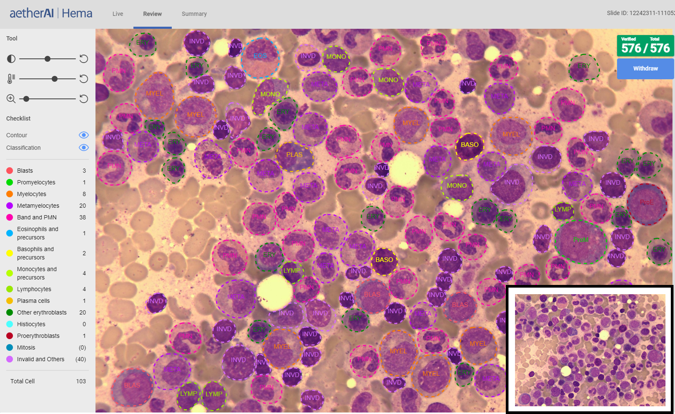
Figure 1. A screen view of the system’s interpretation results.
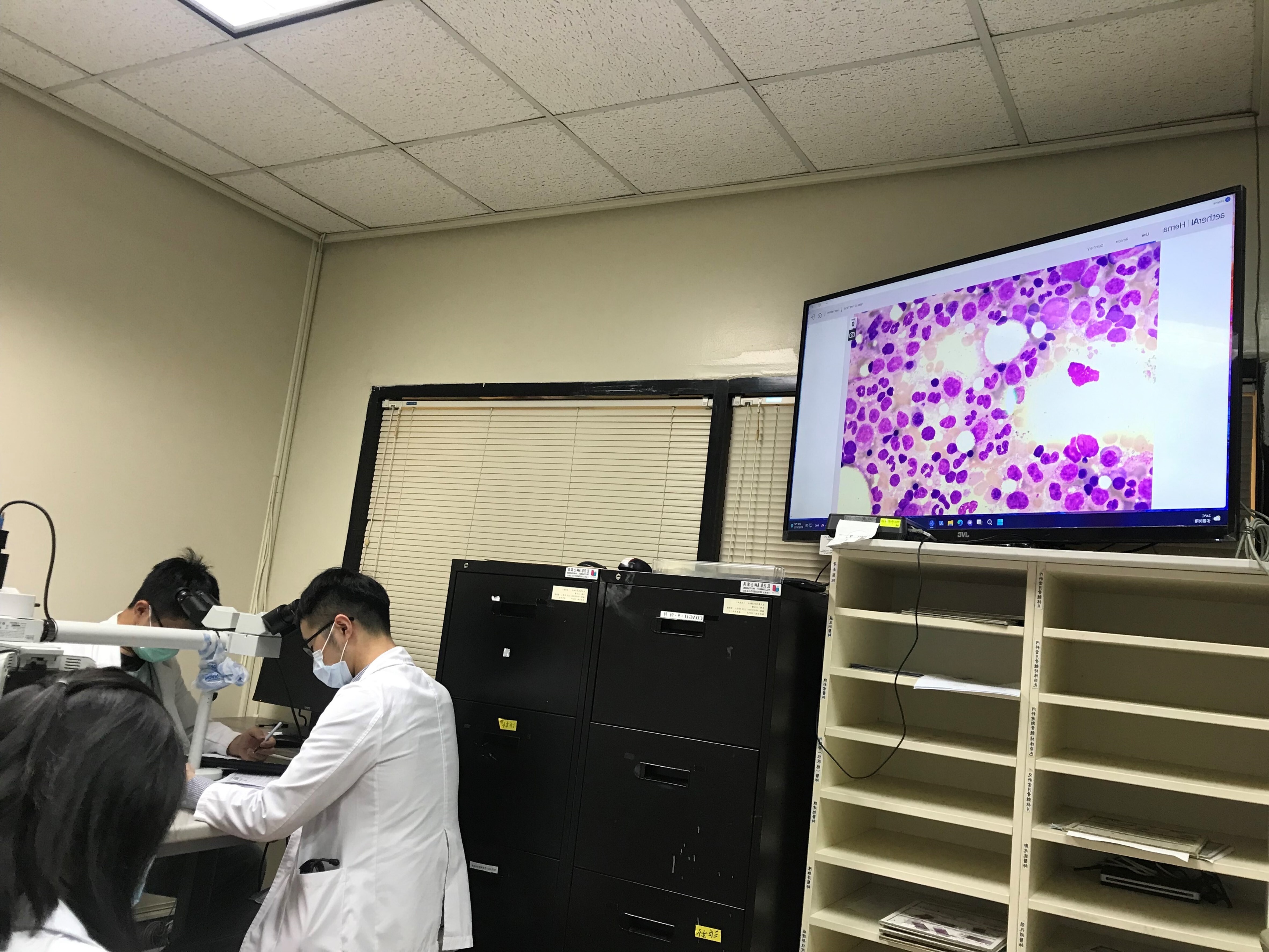
Figure 2. Students learning about the digital pathology system in class.